Bone Cancer
Learn more about bone cancer and how it is treated.
Overview
Bone sarcomas are a rare type of cancer that starts in the cells of the bone or cartilage.
Sarcomas are primary bone cancers. This means they begin in the bone, as opposed to cancers that have spread to the bones from somewhere else in the body.
Types of bone cancers
Osteosarcoma
Osteosarcoma is the most common bone cancer. It is more common in young adults and children, but it can develop in patients of any age.
Osteosarcoma usually forms in the end of the thigh bone (the femur), the top of the shinbone (the tibia), or the upper arm bone (humerus), though it can start in any bone. In older adults, osteosarcoma is often found in the hips and jaws.
Chondrosarcoma
Chondosarcoma is the second most common type of primary bone cancer. It is more common in adults. Chondosarcoma starts in the cartilage, which is the tissue that lines the joints. It is most often found in the upper arm bone (humerus), thigh bone (femur) and pelvic bones.
Ewing sarcoma
Unlike other types of sarcomas, Ewing sarcoma is a group of tumours that can develop in both the bone and the soft tissues. It is most common in young adults, but it can develop in patients of any age.
Ewing sarcoma can begin in the pelvis, the long bones of the arms and legs, the chest (ribs and shoulder blades) and spine.
Diagnosis
There are several tests used to diagnose bone cancer and more than one test is often needed to make an accurate diagnosis.
X-ray
X-rays use a type of radiation called electromagnetic waves to create images of your bones and other structures inside the body. These images help physicians see if there are any abnormalities.
Computed tomography (CT) scan
A CT scan takes pictures of the inside of the body using X-rays from different angles. A computer combines these images into a cross-sectional picture that makes it easier to see if there are any abnormalities or tumours.
CT scan images provide more detailed information than regular X-rays.
Magnetic resonance imaging (MRI)
MRIs use magnet and radio waves to create pictures of your bones. This helps physicians see if there are any abnormalities.
Bone scan
A bone scan consists of both an injection and a scan. A very small amount of radioactive substance is injected into your veins. These substances are called tracers.
After being injected with the tracers, we scan your body with a special camera that picks up on the tracer. The camera is connected to a computer than creates an image of your bones. Changing cells absorb more of the tracer substance, which makes it easier to see if there are any abnormalities.
Positron emission tomography (PET) scan
PET scans are used to look for abnormalities. A very small amount of radioactive substance is injected into your veins. These substances are called tracers.
After being injected with the tracer, you lie down on the PET scanner. The PET scanner creates an image of the inside of your body. Changing cells absorb more of the tracer substance, which makes it easier for physicians to see if there are any abnormalities.
Biopsy
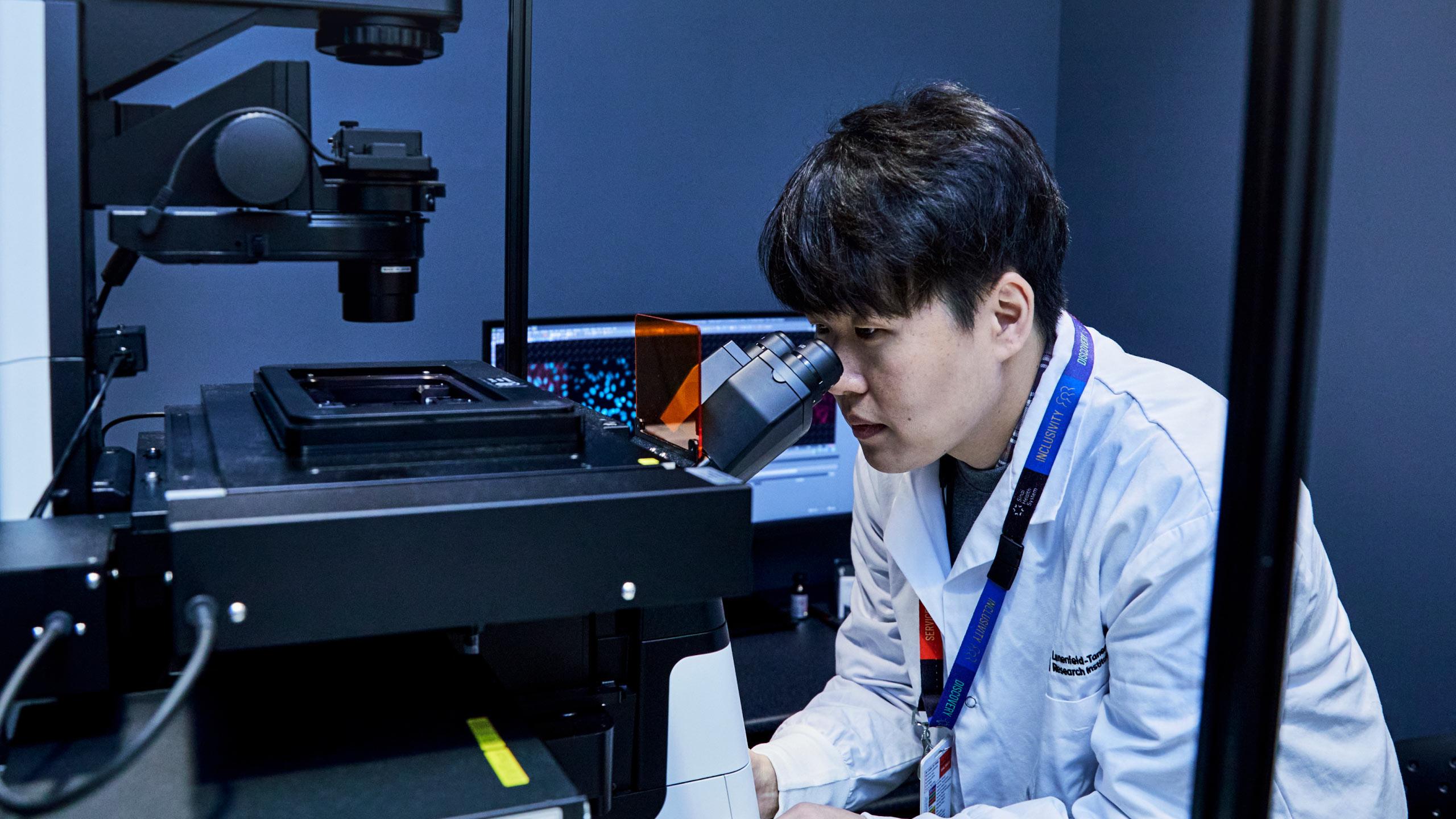
If your imaging test suggests that cancer is present, we need to take a biopsy to make a definite diagnosis. During a biopsy, a physician removes a small amount of tissue so it can be examined under a microscope and analyzed by a pathologist. A pathologist is a physician who specializes in interpreting laboratory tests and evaluating cells, tissues, and organs to diagnose disease.
A bone biopsy requires careful planning and is only performed by sarcoma specialists who have extensive expertise in diagnosing and treating bone cancers.
Treatment
Our sarcoma care team works with you to develop a treatment plan that is tailored to your needs. Your treatment options depend on factors such as the type of sarcoma and the stage of your cancer.
We also consider possible side effects, your overall health and your personal preferences. Part of your treatment may focus on treating cancer symptoms or side effects of treatment that affect your quality of life.
We may recommend one or more of the following treatments.
Surgery
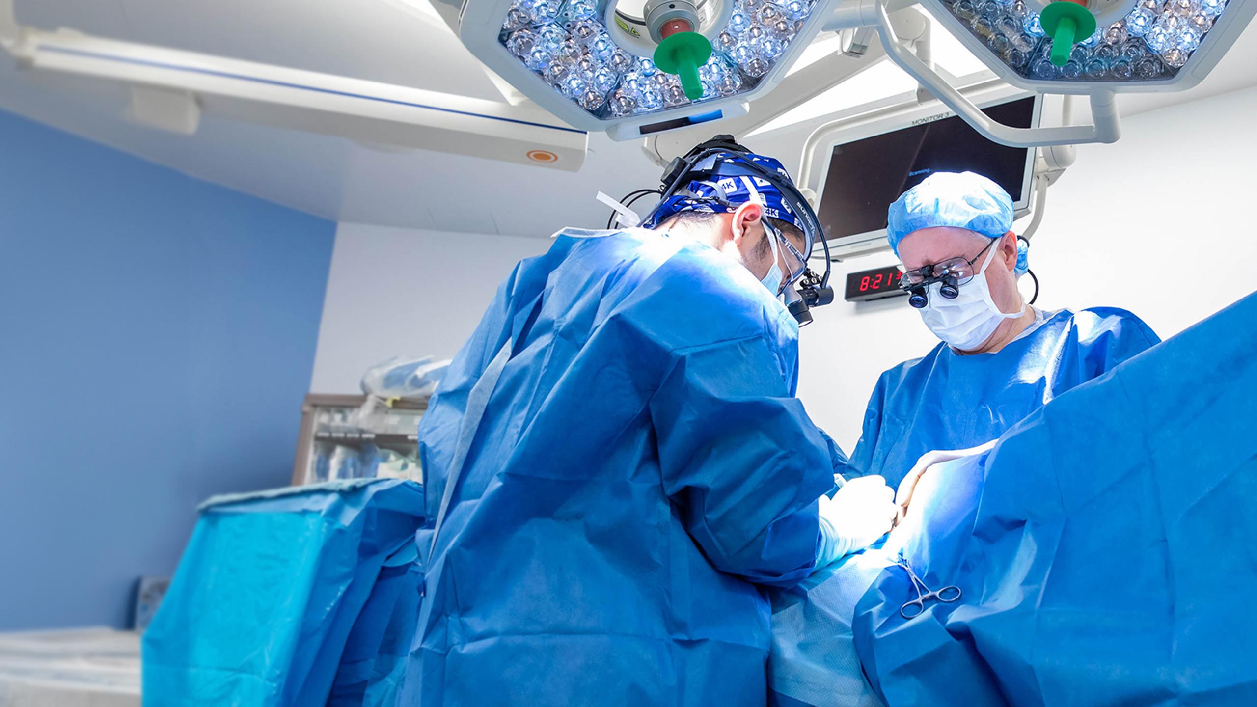
During surgery, an orthopaedic oncologist tries to remove the tumour and some of the healthy bone or tissue around the tumour. This helps remove all the cancer cells. Orthopaedic oncologists are physicians who specialize in treating bone cancer with surgery.
Surgical removal of the tumour is the main treatment for chondrosarcoma since chondrosarcoma does not usually respond well to chemotherapy or radiation therapy.
The following types of surgeries may be used to treat bone cancer.
Wide excision surgery
During wide excision surgery, an orthopaedic oncologist removes the tumour with a margin of healthy bone or tissue around the tumour to ensure all cancer cells have been removed.
Limb-sparing surgery
During a limb-sparing surgery, an orthopaedic oncologist removes the tumour from your arm or a leg while preserving nearby vessels and nerves to avoid removing the whole arm or leg.
Reconstructive surgery
When performing reconstructive surgery, an orthopaedic oncologist replaces the bone that has been removed with prostheses such as metal plates, bone grafts or modular implants (a medical device used to restore mobility). These prostheses help provide strength to the remaining bone.
Muscle flaps and skin grafts may be used over the area that has been reconstructed to help with healing and to reduce the risk of infection.
Amputation
Amputation may be the best treatment option in cases where the tumour cannot be completely removed with surgery or when reconstructive surgery is not possible. Amputation may also be necessary if the cancer returns to the same area after limb-sparing surgery.
Chemotherapy
Chemotherapy is prescribed by a medical oncologist, who is a physician who specializes in treating cancer with medication. It may be given alone or in combination with surgery and/or radiation therapy.
Chemotherapy is more effective in treating some bone cancers than others. Chemotherapy is an important part of the treatment plan for osteosarcoma and Ewing sarcoma, but is rarely effective in the treatment of chondrosarcoma.
Our medical oncologists specialize in the treatment of sarcoma using medication. All bone cancers are unique and require different treatment strategies and may include the use of two or three different chemotherapy drugs.
For fast-growing tumours, you may need chemotherapy before your surgery. Chemotherapy helps shrink the tumour, which makes it easier to remove. This is called neoadjuvant chemotherapy.
After you have recovered from surgery, you may need more chemotherapy to destroy any remaining cancer cells. This is called adjuvant chemotherapy.
The side effects of chemotherapy depend on the type and dose of drugs that are used and your personal reaction to them. Some common side effects include fatigue, low blood counts, risk of infection, nausea and vomiting, hair loss, loss of appetite and diarrhea. These side effects usually go away after treatment is finished. Our care team will monitor you for potential long-term side effects.
For most chemotherapy regimens to treat primary bone cancer, treatments take two to five days. In this case, you need to be admitted to the hospital to receive your chemotherapy. This is called inpatient chemotherapy.
Radiation
Radiation therapy is prescribed by a radiation oncologist. Radiation is generally given over several consecutive days.
Targeted therapy
Targeted therapy uses medicine to target the proteins and genes in the tumour that cause the cancer to grow. This medication blocks the growth and spread of cancer cells and limit damage to healthy cells. Your cancer cells may be tested to see if they respond to targeted therapy.
Immunotherapy
Immunotherapy, also called biologic therapy, is a treatment that uses your immune system to fight the cancer. Your cancer cells may produce proteins that prevent the immune system from attacking them. Immunotherapy medications interfere with that process to target, improve or restore the functioning of your immune system.
