


How award-winning research trainees are transforming our understanding of tumour biology
Most solid cancers remain notoriously difficult to treat and research, largely because no two tumors are alike.
The entanglement of different types of cancer and healthy cells complicates the identification of specific cells and genes driving the disease. This complexity also impedes predictions about cancer progression and response to treatments.
Despite these challenges, researchers at the Lunenfeld-Tanenbaum Research Institute of Sinai Health are confronting tumor heterogeneity head-on. They are pioneering new genomic engineering and tissue profiling to understand the genetic and cellular diversity in tumors, providing valuable insights that could revolutionize therapy.
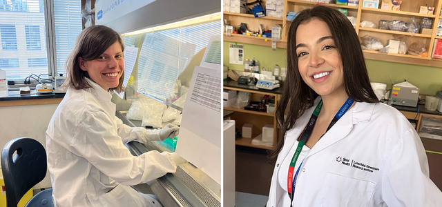
Earlier this year, the Canadian Cancer Society recognized the significance of this work by awarding its inaugural Postdoctoral Fellowships to two outstanding LTRI scientists. Dr. Ellen Langille received an award for her project aimed at identifying genes that cause breast tumors to metastasize to different parts of the body, while Dr. Noor Shakfa focuses on mapping cellular diversity in pancreatic tumors to enhance future immunotherapy approaches.
Deciphering Cancer’s Genetic Puzzle
Each tumor consists of a diverse mix of genetically distinct cancer cells, often containing hundreds of mutations. This diversity makes it challenging to determine which mutations are actually driving the cancer, as many are benign.
Dr. Langille is using a powerful approach to simultaneously examine over a hundred genes in living animals to identify those that promote cancer. This method, developed by Dr. Langille’s supervisor, Dr. Daniel Schramek, a Senior Investigator at LTRI and Deputy Director of Discovery Research for Sinai Health, uses the CRISPR genome editing tool to target specific genes in individual cells in mice. The approach also captures the influence of the microenvironment on cancer development that cannot be modeled in a Petri dish. Ultimately, Dr. Langille can pinpoint critical genes for tumour growth, paving the way for new therapeutic targets.
Dr. Langille completed her PhD with Dr. Schramek during which she identified novel genes that are critical for the development of breast cancer. Intriguingly, she found a link between the normal process of cell proliferation, which occurs in the breast as it prepares for milk production, and cancer. For this work she received the Barbara Vivash Award in Molecular Genetics that recognizes the best PhD thesis completed in the Department of Molecular Genetics at the University of Toronto. Enticed by the technology’s potential, Dr. Langille decided to stay in the lab and apply the approach to identifying the genes in cancer cells, and in their healthy neighbours, that enable breast tumours to spread to other parts of the body.
“Daniel has been an incredibly supportive mentor. He opened many doors for me during my PhD and connected me with researchers with expertise outside our lab that expanded my horizons,” said Dr. Langille. “Winning this fellowship was a significant validation, affirming that others also value our research.”
Dr. Langille’s award is co-supported by the Canadian Institutes of Health Research
Mapping cellular diversity in pancreatic tumours
Dr. Noor Shakfa is also taking advantage of specialized technology to analyze the cellular makeup of pancreatic tumors, known for their heterogeneity and treatment resistance. Her trailblazing project aims to produce the most detailed spatial map yet of the different cancer cells, immune cells, fibroblasts and other cell types that make up these tumors, providing key insights for targeted immunotherapy. Immunotherapy, where patient’s own immune system is harnessed for fighting cancer, offers great promise but currently benefits only a subset of cancers and patients. Dr. Shakfa and scientists like her believe that progress can be made by understanding the specific local cellular interactions within tumors.
She joined the laboratory of Dr. Hartland Jackson, an Investigator at the LTRI and a leading expert in a technology called Imaging Mass Cytometry (IMC), which allows for detailed spatial examination at the subcellular level within intact tissues. This is critical for understanding interactions and relationships between cells, as these disappear when cells are placed in suspension for conventional analyses. With IMC, high resolution images of patient tumors and computational analysis of single cells can be explored to build detailed landscapes of tumour cells in their environment.
Dr. Shakfa, who investigated tumour-immune interactions in ovarian cancer during her PhD at Queen’s University, was drawn to join Dr. Jackson’s lab by his expertise and the advanced resources available at LTRI—including Canada’s first whole-tissue imaging system and comprehensive computational analysis pipelines. This setup enables high-throughput collection and analysis of valuable patient samples.
“I am immensely grateful for Hart’s support and access to top-notch technology and resources,” Dr. Shakfa expressed. “Receiving the CCS fellowship, especially knowing it was partly adjudicated by patients, has been incredibly humbling and motivating.”
Dr. Shakfa’s award is co-supported by the Terry Fox Research Institute.
Related news and stories
View all News and Stories


